Diagnostic Imaging offers a comprehensive suite of breast screening and diagnostic services, from an initial baseline screening mammogram to diagnostic examinations and ultrasound-guided biopsy, all in one convenient location.
All women are unique, so when it comes to breast imaging there is no “one-size-fits-all” approach. We’ll work with you and your doctor to make sure you receive the care you need at all of life’s stages. We maintain the latest 3D mammography and ultrasound technology, and our radiologists offer unparalleled expertise in breast imaging.
Mammography
Mammograms detect breast abnormalities using low-dose x-rays. Mammograms help radiologists to detect breast cancer, cysts or calcifications, usually before they can even be felt during a breast exam.
There are two types of mammograms:
- A screening mammograms aids in the early detection of breast cancer and is recommended for women aged 40 and older who show no symptoms of a breast abnormality. If a woman is at high risk, she should talk to her doctor about starting screening mammograms earlier than age 40.
- A diagnostic mammogram is used to evaluate a specific area of the breast when a potential abnormality is found.

Convenient Care and a Personal Touch
Ask your provider to refer you to Diagnostic Imaging.
Why have your mammogram at Diagnostic Imaging?
All mammograms at our center are performed on the Hologic Selena 3D mammography system. It’s the most advanced form of mammography, also known as digital breast tomosynthesis. It can detect smaller cancers that may be missed on traditional 2D mammography.
At Diagnostic Imaging, all patients receive a 3D screening mammogram; there is no extra upcharge like there is with other centers. In addition, we will schedule your mammogram quickly, in many cases on the same day or within a few days of your request. We accept all insurance, including Medicare and Medicaid. Finally, all screening mammogram reports are provided to you and your doctor in 3 days or less, so there is no needless waiting for test results. Diagnostic mammogram reports are delivered within 24 hours of your test. If you are uninsured, call us! Our cash price for screening mammograms is very reasonable and affordable.

Mammography at Diagnostic Imaging is accredited by the American College of Radiology
Breast Ultrasound
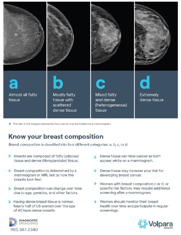
If an abnormality is found on a mammogram, or if a mammogram is not conclusive, a breast ultrasound may be ordered by your doctor. There are other reasons for an ultrasound to be suggested, such as if a woman has dense breasts, is very young or if she is pregnant.
Ultrasound produces images of the breast using high-frequency sound waves. As the sound waves bounce off internal breast tissues, they create echoes which a computer translates into images on a screen. Like other imaging tests, breast ultrasound can show abnormalities and disease within the breast. It is fast, painless and completely free of radiation or harmful side effects. Breast ultrasound is also used to determine if an abnormality is a non-cancerous (benign) or cancerous (malignant) tumor. This can help you potentially avoid a breast biopsy.
Ultrasound-guided biopsy
If a biopsy is required, Diagnostic Imaging performs ultrasound-guided biopsy. This non-surgical option is less invasive and precise. During a biopsy, a core sample is taken from a suspicious area and sent to a laboratory for examination to confirm or rule out a cancer diagnosis.
What to Expect
On the day of your appointment, don’t use deodorant, powders, ointment or perfume on your breasts or underarm area. These products can contain ingredients that may interfere with the quality of images. You will be asked to undress from the waist up and you will be given a gown to wear.
Mammograms
A technologist will position your breast between two plates, which will compress the breast so as much breast tissue as possible can be imaged. The technologist will then reposition your breast on the plates to be imaged from a different angle. The process is repeated on the other side.
If you’ve had prior mammograms at facilities other than Diagnostic Imaging, please request the images be sent to our office prior to your appointment or bring them with you. The ability to compare current images to prior images allows radiologists to see subtle changes over time, which is helpful in detecting cancers at their earliest stages.
The results of your mammogram are communicated to your referring provider after your exam. If you have questions about your results, you are encouraged to speak with your referring provider, who can discuss your results in detail with you.
If you would like to use the Tyrer-Cuzick Risk Assessment Calculator, (please click here) this calculator asks questions about your personal and family history to determine your lifetime risk probability of developing breast cancer compared with the age-adjusted U.S. population average probability of developing breast cancer. The purpose of this tool is simply to inform you. Please consult with your healthcare provider should you have any questions about your risk of developing breast cancer or for guidance on options for breast cancer screening or genetic counseling.
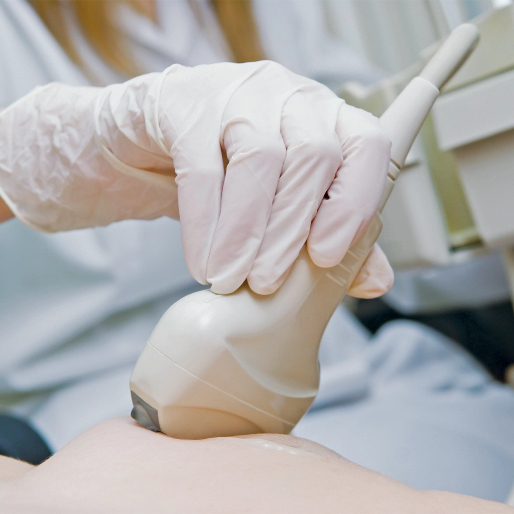
Breast ultrasound and ultrasound-guided biopsy
During a breast ultrasound, the technologist will move a wand-like device called a transducer over the skin on your breast. They will press the transducer firmly across the area of your breast being imaged. The images are then recorded and will be read at a later time by the radiologist.
There are no diet restrictions prior to an ultrasound so you may eat, drink and take medications as you normally would. Please wear clothing that is easy to take off or that easily enables the technologist to reach your chest. A special gel is used on the transducer. It does not stain clothing, but it is sticky so there may be some residue on your skin after your ultrasound examination.
If you are having an ultrasound-guided biopsy, you may be asked to wear or bring a tight-fitting brassiere or sports bra. This can help to minimize breast motion and potential discomfort.